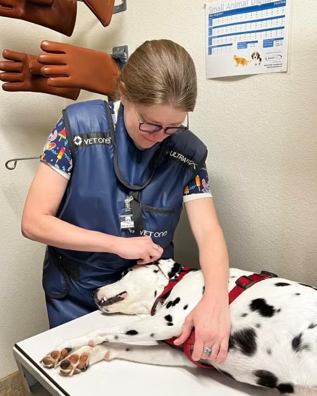
PennHIP Method: Advancing Hip Joint Assessment for Canine Health
 The PennHIP method introduces a groundbreaking approach to evaluating, measuring, and interpreting hip joint laxity in canines. Comprising three distinct radiographs - the distraction view, the compression view, and the hip-extended view - this innovative technique has revolutionized the field of hip screening and has demonstrated superior accuracy compared to the conventional standard.
In the realm of canine hip assessment, precision is paramount. The PennHIP method utilizes the distraction view to achieve precise measurements of joint laxity, while the compression view captures the critical aspect of joint congruity. By obtaining comprehensive data on both laxity and congruity, this method provides a comprehensive understanding of hip joint health. In addition, the hip-extended view offers supplementary insights into the presence of osteoarthritis (OA) within the hip joint. Unlike the traditional, subjectively scored hip-extended radiograph often referred to as the OFA view, the PennHIP technique offers a more accurate and reliable means of predicting the onset of OA.
Ensuring optimal diagnostic radiographs necessitates the complete relaxation of the patient and its surrounding musculature. Achieving this state of relaxation requires the administration of sedation and/or general anesthesia, prioritizing the comfort and safety of the animal. Typically, the evaluation process involves capturing three separate radiographs. The compression view involves positioning the femurs neutrally and pushing the femoral heads fully into the sockets. This positioning provides a true depiction of the hip socket's depth and the ball-and-socket fit. The subsequent distraction view maintains a neutral orientation while applying a controlled lateral force through a specialized positioning device. This position, renowned for its accuracy and sensitivity, reveals the extent of passive hip laxity. Studies have consistently shown that passive hip laxity is a key risk factor in the development of hip dysplasia-associated osteoarthritis. The final component, the hip-extended view, facilitates the assessment of pre-existing joint diseases, such as osteoarthritis.
One notable hallmark of the PennHIP technique is its versatility in application. Studies conducted by PennHIP have extensively examined the method's efficacy, spanning from eight weeks to three years of age. Impressively, the PennHIP method can reliably be employed on puppies as young as 16 weeks old. Notably, the passive hip laxity observed at 16 weeks demonstrates a strong correlation with later hip laxity, indicating that a dog's hip condition at 16 weeks tends to persist throughout its development. German Shepherd Dogs showcase particularly consistent results in this regard. However, the accuracy of laxity measurements for breeds other than German Shepherds is still under scrutiny, with further research needed to determine the earliest reliable age of evaluation.
In conclusion, the PennHIP method stands as a remarkable advancement in the realm of canine health assessment. Its innovative approach, coupled with its accuracy in predicting hip joint health and potential osteoarthritis, has revolutionized hip screening practices. By embracing the PennHIP method, veterinarians and dog owners alike can proactively manage canine hip health, enhancing the quality of life for our four-legged companions.
